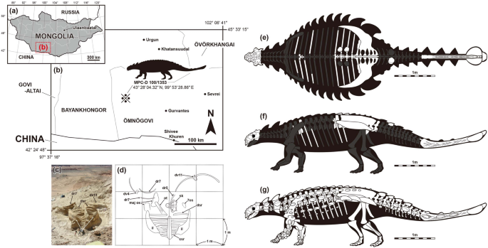
A new ankylosaurid from the Upper Cretaceous Nemegt Formation of Mongolia and implications for paleoecology of armoured dinosaurs A new ankylosaurid dinosaur, Tarchia tumanovae sp. nov., has been recovered from the Upper Cretaceous Nemegt Formation of Mongolia. It includes a well-preserved skull, dorsal, sacral, caudal vertebrae, sixteen dorsal ribs, ilia, a partial ischium, free osteoderms, and a tail club. The squamosal horns of T. tumanovae are divided into two layers, the external dermal layer and the underlying squamosal horn proper. The irregular ventral margin of the base of the upper dermal layer may represent a resorption surface, suggesting that the squamosal horns of some ankylosaurids underwent extreme ontogenetic remodeling. Localized pathologies on the dorsosacral ribs and the tail provide evidence of agonistic behaviour. The tail club knob asymmetry of T. tumanovae resulted from restricted bone growth due to tail club strikes. Furthermore, T. tumanovae had an anteriorly protruded shovel-shaped beak, which is a morphological character of selective feeders. Ankylosaurid diets shifted from low-level bulk feeding to selective feeding during the Baruungoyot and the Nemegt ‘age’ (middle Campanian-lower Maastrichtian). This ankylosaurid niche shifting might have been a response to habitat change and competition with other bulk-feeding herbivores. Ankylosaurid dinosaurs, one group of Ankylosauria, are quadrupedal, herbivorous, and have a heavily ornamented skull and parasagittal rows of osteoderms covering the dorsolateral surfaces of the body1. Their fossils have been frequently discovered from the Upper Cretaceous (upper Campanian-lower Maastrichtian) Nemegt Formation of Mongolia2,3,4,5,6,7,8,9. Except for the holotype of Tarchia teresae (PIN 3142/250), previously known specimens from this rock unit consist of only postcranial material, mostly caudal elements.During the Korea-Mongolia International Dinosaur Expedition in 2008, a new skull with a partial postcranial skeleton (MPC-D 100/1353) was collected from the Nemegt Formation in Hermiin Tsav (Fig. 1). The specimen includes a well-preserved skull, dorsal, sacral, caudal vertebrae, sixteen dorsal ribs, ilia, a partial ischium, free osteoderms, and a tail club (Figs. 2, 3, 4, 5, 6, 7, 8), and turned out to be a new taxon. As a new taxon, it is named, described, and discussed herein. The new specimen provides further evidence of ontogeny, agonistic behavior, and suggestions of niche shifting in Mongolian ankylosaurids.Figure 1Map showing the locality where Tarchia tumanovae sp. nov. (MPC-D 100/1353) was discovered (a–d). (a) Map of Mongolia. (b) Enlarged map of the dotted lined rectangle of A marked with the fossil locality (※). (c) Photo of the excavation site. (d) Quarry map showing bone location. (e–f) Skeletal diagram of the specimen in dorsal (e) and left lateral (f) views (white bones represent recovered elements). (g) Skeletal reconstruction with dermal armour. Abbreviations: 7os, Type 7 osteoderm; csr, caudosacral vertebra; dr, dorsal rib; dsr, dorsosacral vertebra; il, ilium; maj os, major osteoderm; ot, ossified tendon; sk, skull. Adobe Illustrator CC (version 24.0.1, https://www.adobe.com/kr/products/illustrator.html) was employed to produce (a–g).Figure 2Photographs (a–d) and line drawings (e–h) of the skull of Tarchia tumanovae sp. nov. (MPC-D 100/1353). Photographs of the skull in (a) left lateral, (b) right lateral, (c) anterior, and (d) occipital views. Line drawings in (e) left lateral, (f) right lateral, (g) anterior, and (h) occipital views. Grey areas indicate damaged surfaces. apt A aperture A, apt C aperture C, asob anterior supraorbital caputegulum, fm foramen magnum, gr groove, inca internarial caputegulum, laca lacrimal caputegulum, lnca lateral nuchal caputegulum, loca loreal caputegulum, mnca medial nuchal caputegulum, mx maxilla, naca nasal caputegulum, oc occipital condyle, orb orbit, parocc paroccipital process, pmx premaxilla, pmxo premaxillary ornamentation, prfca prefrontal caputegulum, prot protuberance, psob posterior supraorbital caputegulum, pt pterygoid, q quadrate, qjh quadratojugal horn, sn n supranarial notch, snca supranarial caputegulum, sp small process between the foramen magnum and the nuchal shelf, sqh squamosal horn, v vomer. Adobe Illustrator CC (version 24.0.1, https://www.adobe.com/kr/products/illustrator.html) was employed to produce (e–h).Figure 3Photographs (a and b) and line drawings (c and d) of the skull, and photographs of the maxillary teeth (e–g) of Tarchia tumanovae sp. nov. (MPC-D 100/1353). Photographs of the skull in (a) dorsal and (b) palatal views. Line drawings in (c) dorsal and (d) palatal views. Grey areas indicate damaged surfaces, and solid diagonal lines indicate unremoved matrix. Photographs of (e) seventh left, (f) third right, and (g) eight right maxillary teeth in anterolabial view. anca anterolateral nuchal caputegulum, asob anterior supraorbital caputegulum, bas basioccipital, bas fo basioccipital foramen, bs basisphenoid, c cingulum, d denticle, ect ectopterygoid, fca frontal caputegulum, inca internarial caputegulum, laca lacrimal caputegulum, lnca lateral nuchal caputegulum, loca loreal caputegulum, mnca medial nuchal caputegulum, mso middle supraorbital caputegulae, mx maxilla, naca nasal caputegulum, oc occipital condyle, pal palatine, parocc paroccipital process, pca parietal caputegulum, pmx premaxilla, pmxo premaxillary ornamentation, prfca prefrontal caputegulum, psob posterior supraorbital caputegulum, pt pterygoid, pt fo pterygoid foramen, q quadrate, qjh quadratojugal horn, snca supranarial caputegulum, sqh squamosal horn, v vomer. Adobe Illustrator CC (version 24.0.1, https://www.adobe.com/kr/products/illustrator.html) was employed to produce (c and d).Figure 4Photographs of dorsal vertebrae of Tarchia tumanovae sp. nov. (MPC-D 100/1353). The fourth dorsal vertebra in (a) left lateral, (b) right lateral, (c) dorsal, and (d) ventral views. Fourth dorsal vertebra with fused right rib in (e) anterior and (f) posterior views. The eleventh dorsal vertebra in (g) anterior, (h) posterior, (i) left lateral (with no ribs attached), and (j) ventral views. dr dorsal rib, nc neural canal, ns neural spine, pa parapophysis; prezygapophysis, poz postzygapophysis, tp transverse process.Figure 5Photographs of dorsal (a–h) and dorsosacral ribs (i–p) of Tarchia tumanovae sp. nov. (MPC-D 100/1353). Both third dorsal ribs in (a) anterior and (b) posterior views. The fourth left dorsal rib in (c) anterior and (d) posterior views. The fifth left dorsal rib in (e) anterior and (f) posterior views. The sixth left dorsal rib in (g) anterior and (h) posterior views. Both first dorsosacral ribs in (i) anterior and (j) posterior views. Both second dorsosacral ribs in (k) anterior and (l) posterior views. Both third dorsosacral ribs in (m) anterior and (n) posterior views. ca capitulum, path pathology, tu tuberculum.Figure 6Photographs of the synsacrum (a–f), and ilia (g) of Tarchia tumanovae sp. nov. (MPC-D 100/1353). Synsacrum in (a) left lateral, (b) right lateral, (c) ventral, (d) anterior, and (e) posterior views. (f) Second caudosacral vertebra in left lateral view. (g) Synsacrum with the fused ilia in dorsal view. cs caudosacral vertebra, csr caudosacral rib, ds dorsosacral vertebra, dsr dorsosacral rib, il ilium, ns neural spine, psr parasacral rib, psv parasacral vertebra, sr sacral rib, sv sacral vertebra.Figure 7Photographs of both ilia (a–d), partial left ischium (e–g), and the tail club (h–k) of Tarchia tumanovae sp. nov. (MPC-D 100/1353). (a) Left and (b) right ilium in ventral view. (c) Right and (d) left ilium in lateral view. Left ischium in (e) lateral, (f) medial, and (g) proximal views. Tail club handle in (h) left lateral and (i) right lateral views. (j) Tail club handle and knob in dorsal view. (k) Tail club knob in proximal view. ac acetabulum, ch chevron, csr caudosacral rib, maj os major osteoderm, min os minor osteoderm, ns neural spine, path pathology, poa postacetabular process, prea preacetabular process, psr parasacral rib, sr sacral rib.Figure 8Photographs of ossified tendons from the tail knob handle (a–d) and dermal osteoderms (e–m) of Tarchia tumanovae sp. nov. (MPC-D 100/1353). (a) A bundle of ossified tendons from the tail knob handle. Close up photos of (b) an ossified tendon that is flattened and (c) a tendon that is elliptic in cross-section and has longitudinal striae. (d) An ossified tendon with evidence of fracture healing. Type 2 osteoderm in (e) anterior, (f) oblique dorsolateral, and (g) ventral views. Two Type 7 osteoderms in (h and k) dorsal, (i and l) ventral, and (j and m) lateral views. k keel, path pathology.Dinosauria Owen10.Ankylosauridae Brown11.Ankylosaurinae Nopcsa12.Tarchia Maryañska13.Type species. Tarchia kielanae Maryañska13.Revised diagnosis. An ankylosaurid distinguished by having the following unique set of characters (autapomorphies with an asterisk): a narrow internarial bar of the premaxillae (shared with Tsagantegia) (ambiguous in Tarchia kielanae); large, rhomboidal loreal caputegulum with a laterally extended posterior keel (shared with Saichania) (ambiguous in T. kielanae); subrectangular frontal caputegulae (shared with Saichania); a ‘neck’ present at the base of the quadratojugal horn (shared with Pinacosaurus mephistocephalus and Minotaurasaurus) (ambiguous in T. kielanae); sigmoidal and peaked anteromedial supraorbital caputegulum*; posterolateral supraorbital caputegulum with a rounded anterior surface, and a flat, anteriorly-inclined posterior surface*; anteromedially poorly defined postorbital fossa that medially reaches the lateral nuchal caputegulae*; occiput visible in dorsal view (shared with Minotaurasaurus and Zaraapelta); foramen magnum taller than wide*. Differs from Minotaurasaurus, Pinacosaurus grangeri, Saichania, and Zaraapelta in having no postocular caputegulae (ambiguous in T. kielanae) and a posteroventrally oriented occipital condyle. Differs from Minotaurasaurus, P. grangeri, and Zaraapelta in having confluent supraorbital horns. Differs from Minotaurasaurus and Saichania in having a relatively tall braincase. Differs from Minotaurasaurus and Zaraapelta in having a long nuchal crest. Differs from Minotaurasaurus in having relatively long paroccipital processes that laterally reach the squamosal horns. Differs from Saichania in having remodeled squamosal horns and anteroposteriorly short lateral nuchal caputegulae.Tarchia tumanovae sp. nov.Etymology. Named in honour of Tatiana Tumanova for her contributions toward the understanding of Mongolian ankylosaurs.Holotype. MPC-D 100/1353 (Figs. 1, 2, 3, 4, 5, 6, 7, 8), a well-preserved skull, dorsal, sacral, caudal vertebrae, sixteen dorsal ribs, ilia, a partial ischium, free osteoderms, and tail club.Locality and horizon. Upper Cretaceous (upper Campanian-lower Maastrichtian) Nemegt Formation, Hermiin Tsav, southern Gobi Desert, Mongolia.Diagnosis. An ankylosaurid distinguished by having the following unique set of characters: a single relatively bulbous internarial caputegulum that does not reach the rostral tip of beak*; a nasofrontal sagittal furrow with a weak Z-shaped offset (shared with Tarchia kielanae); lateral nuchal caputegulae taller laterally than medially (shared with Saichania); vomerine keel extends below the alveolar ridge (shared with Saichania). Differs from Minotaurasaurus, Pinacosaurus grangeri, T. kielanae, T. teresae, and Zaraapelta in having a moderate-sized basioccipital foramen. Differs from Minotaurasaurus, P. grangeri, Saichania, and Zaraapelta in having no postocular caputegulae and a posteroventrally oriented occipital condyle. Differs from Minotaurasaurus, P. grangeri, T. teresae, and Zaraapelta in having an anteriorly situated quadrate-quadratojugal region. Differs from Minotaurasaurus, P. grangeri, and Zaraapelta in having confluent supraorbital horns. Differs from P. grangeri, Saichania, and Zaraapelta in having a tall foramen magnum. Differs from Saichania, T. kielanae, and Zaraapelta in having unfused quadrate to the exoccipital area. Differs from Minotaurasaurus and Saichania in having a relatively tall braincase. Differs from Minotaurasaurus and Zaraapelta in having subrectangular frontal caputegulae and a long nuchal crest. Differs from Minotaurasaurus in having narrow narial caputegulae and long paroccipital processes that laterally reach the squamosal horns. Differs from Saichania in having remodeled squamosal horns, anteroposteriorly short lateral nuchal caputegulae, and occiput visible in dorsal view. Differs from T. teresae by having an interpterygoid vacuity visible in occipital view.Description. The shape of the skull is trapezoidal and broader than long in dorsal view (Fig. 3, see Supplementary Information S1 for measurements). All caputegulae are pitted externally. The neuroanatomy of MPC-D 100/1353 was fully described before by Paulina-Carabajal et al.9.The premaxillae are fused dorsally, but the palatal surfaces are separate (Figs. 2, 3). The rostral tip is protruded anteroventrally, and a premaxillary notch is present. The narrow internarial bar is oriented posterodorsally. A thin, rugose ossification is present on each premaxilla below the external naris. The anterior margin of the premaxillary ornamentation is convex, whereas the posterior margin is concave. The palatal surface of the premaxillae is shovel-like with a round anterior boundary. No premaxillary teeth are present. The subcircular external nares face anteriorly. The entrance to the airway (aperture A, sensu14) is large and subcircular. The airway is filled with a matrix. A supranarial notch is present on the medial wall of the maxillary region, lateral to the entrance to the airway. A short, medioventrally sharp intranasal process is present beneath the entrance of this aperture. A larger oval dorsolaterally-facing paranasal aperture (aperture C) is situated on the ventral wall of the external nares behind the internasal bar. The external nares are rimmed dorsomedially, dorsally, and laterally by supranarial caputegulae (sensu15). The dorsomedial and dorsal portion of the supranarial caputegulum is thin, whereas the wide ventrolateral part gives the caputegulum a boot-like appearance in lateral view. The anterior end of the nasals contacts the internasal bar anteromedially. A single medium-sized bulbous internarial caputegulum is situated above the contact between the internarial bar of the premaxillae and the anterior nasals and between the thin medial portion of the two supranarial caputegulae. Behind the internarial caputegulum, eleven pyramidal nasal caputegulae are present. Most of these caputegulae are large and surrounded by a broad sulcus. The most medioposteriorly positioned pair are transversely oriented, pyramidal, and rectangular, similar to the frontal caputegulae.The maxillae are anteroposteriorly elongate, extending to below the orbits. In lateral view, the anterior portion of the maxillae is covered by the loreal caputegulum, whereas the posterior portion is exposed. In palatal view, the convex maxillary tooth row is situated medial to the buccal emargination. Nineteen alveoli are present in each maxilla. A single loreal caputegulum is present on each side.
https://www.nature.com/articles/s41598-021-02273-4
A new ankylosaurid from the Upper Cretaceous Nemegt Formation of Mongolia and implications for paleoecology of armoured dinosaurs